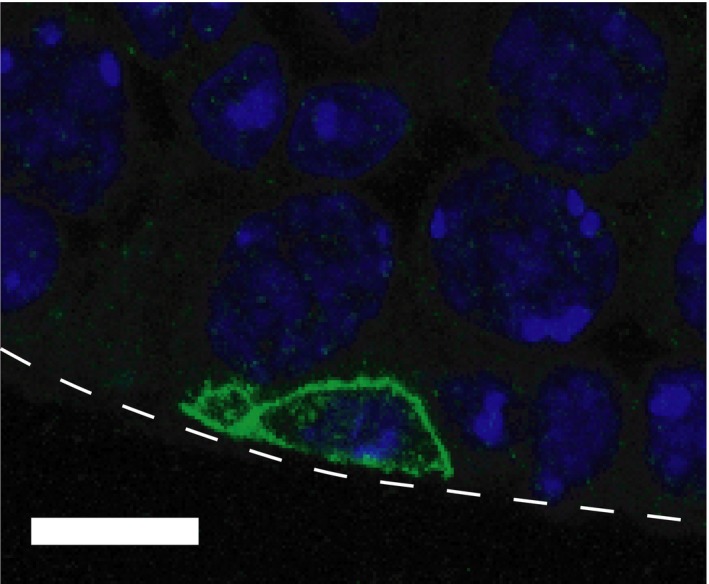
Figure 3.

Undifferentiated spermatogonium on the basement membrane in adult mouse testis. A confocal image of GFRα1+ spermatogonium (green) and nuclear (blue) in C57BL6 adult (3‐mo‐old) mouse testis. GFRα1+ cell was visualized by immunohistochemistry by using anti‐GFRα1 antibody and Alexa488‐conjugated anti‐goat IgG antibody with a nuclear staining by Hoechst 33342, following the procedures described in a previous papers.23, 32 White dotted line indicates a periphery of the seminiferous tubule. Bar indicates 10 μm
