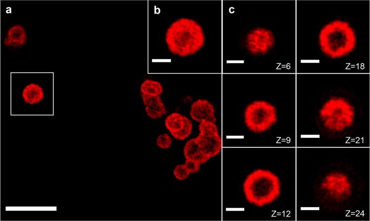
Figure 3.
Characterization of Cy5-labeled hRNFs by SIM imaging. (a) Representative SIM image of Cy5-labeled hRNFs and (b) a higher-magnification view of the particle in the white box in a. (c) Individual frames of the z-stack (step size: 100 nm) of the hRNF particle in b are shown in the right panel. Scale bars: 5 μm (main panel), 1 μm (inset and right panels).

