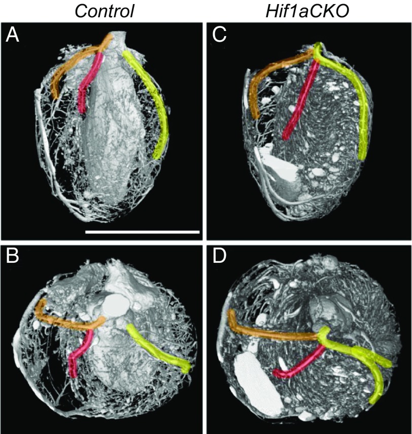
Fig. 5.
Main coronary artery anomalies in Hif1aCKO hearts. Micro-CT visualization of coronary artery branching patterns in 2-mo-old control (A and B) and Hif1aCKO (C and D) mice. Anterior aspect (A and C) and top view (B and D) are shown. Arteries are highlighted from their origin: right coronary artery (RCA; orange), left coronary artery (LCA; yellow), and septal artery (SA; red). The classic arrangement with two independent coronary arteries, the RCA and LCA, arising separately from the aorta and the SA arising from the RCA was observed in 15 out of 15 control hearts (A and B). An anomalous single coronary artery arising from the aorta with subsequent division of the RCA, SA, and LCA was observed in 2 out of 13 Hif1aCKO hearts (C and D). (Scale bar: 5 mm.)

