Fig. 8.
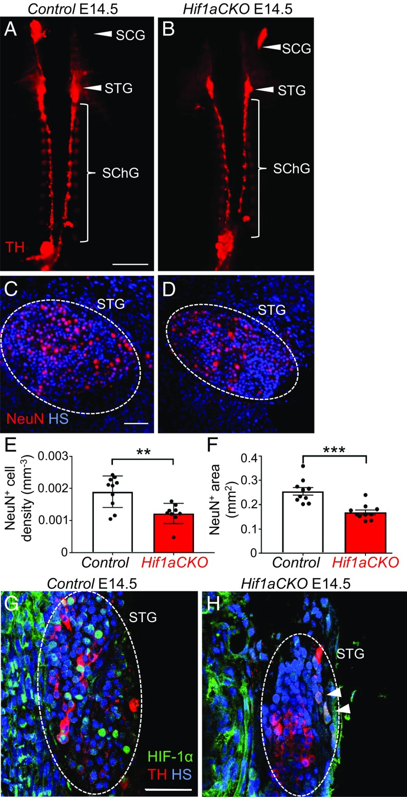
The growth and maturation of secondary sympathetic ganglia are impaired in E14.5 Hif1aCKO embryos. (A and B) Whole-mount TH immunostaining was performed to detect the secondary chain of sympathetic ganglia, which are smaller in Hif1aCKO mice compared with control littermates. Arrowheads indicate the superior cervical (SCG) and stellate (STG) ganglia; bracket indicates the thoracic chain of sympathetic ganglia (SChG). (Scale bar: 1,000 μm.) (C and D) Immunostaining for NeuN (a marker for differentiated neurons; red) shows reduced neuronal density in the STG of Hif1aCKO compared with control embryos (n = 5 embryos per genotype and 2 ganglia per embryo). **P < 0.01, Student’s t test. (Scale bar: 50 μm.) (E and F) Morphometry of sympathetic ganglia (the STG and three upper ganglia of the thoracic sympathetic chain) by whole-mount NeuN immunostaining of the spinal cord (n = 5 embryos per genotype, left and right sympathetic chain ganglia per embryo). ***P < 0.0005, Student’s t test. (G and H) Coronal sections of embryos immunolabeled with anti-TH (red) and anti–HIF-1α (green) show a smaller STG area (dotted oval) and lower HIF-1α expression in Hif1aCKO embryos. Strong HIF-1α expression was detected in nuclei of TH+ neurons in control embryos (G) but only in the cytoplasm of a few TH+ neurons in Hif1aCKO embryos (H, arrowheads). (Scale bar: 50 μm.) All numerical data are presented as mean ± SEM.

