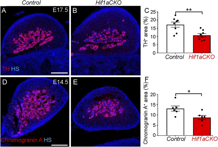
Fig. 9.
Decreased number of adrenal chromaffin cells in Hif1aCKO embryos. (A and B) Cross-sections through the adrenal glands of Hif1aCKO and control embryos at E17.5 were stained with anti-TH antibody (red). (C) TH+ area was quantified using ImageJ software and expressed as percentage of total adrenal gland area (n = 4 embryos per genotype and two sections per embryo). **P < 0.005, Student’s t test. (D and E) Chromogranin A expression in cross-sections of the adrenal glands of control and Hif1aCKO E14.5 embryos was detected by immunostaining. (F) Chromogranin A+ area was quantified using ImageJ and expressed as a percentage of the total adrenal gland area (n = 3 embryos per genotype and two sections per embryo). *P < 0.05, Student’s t test. (Scale bar: 200 μm.) All numerical data are presented as mean ± SEM.

