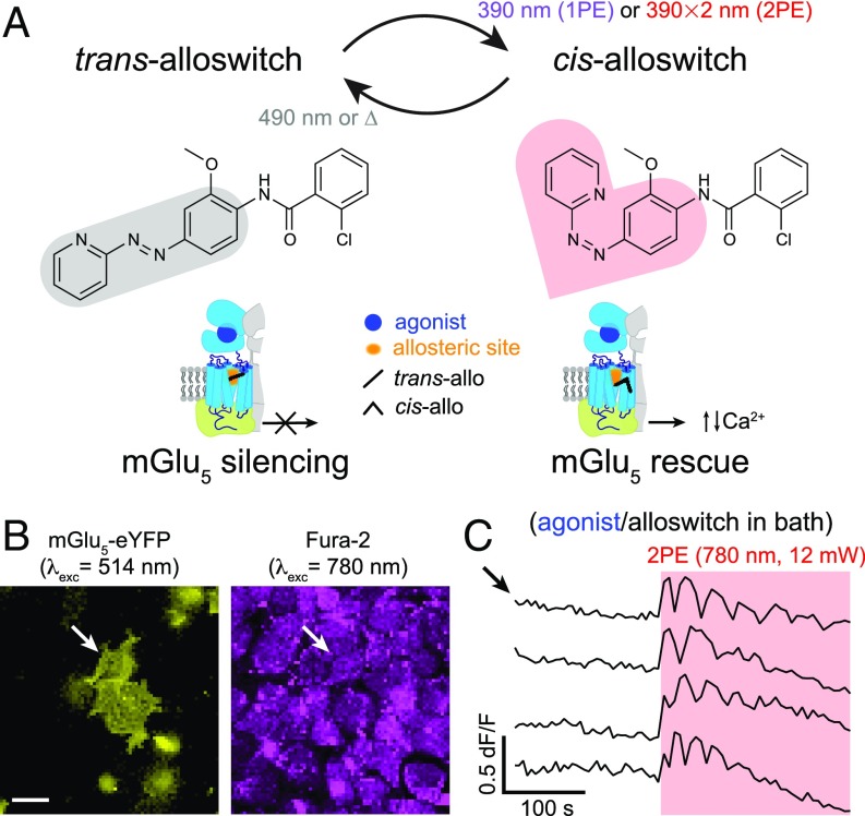
Fig. 1.
Two-photon excitation of alloswitch at NIR wavelengths in cultured cells. (A, Upper) Trans → cis photoisomerization of alloswitch occurs by 1PE under violet light (390 nm). Using pulsed-lasers for 2PE, the expected wavelength for photoswitching is double that required for 1PE. Back-isomerization occurs by thermal relaxation (∆; half-life of cis-isomer, t1/2 = 80 s) (16) or under 490-nm light. The photosensitive core of alloswitch, azobenzene, is highlighted in its trans (gray shade) and cis configurations (red shade). (Lower) Trans-alloswitch silences active, agonist-bound mGlu5 receptors by binding at the allosteric pocket, whereas trans → cis photoisomerization of alloswitch rescues the silenced, agonist-bound receptors at the site of illumination by releasing intracellular, agonist-induced Ca2+ oscillations. (B) HEK cells expressing mGlu5-eYFP (yellow, Left) and loaded with Fura-2 (magenta, Right) for 2-photon imaging of Ca2+ (760–820 nm; shown here is 780 nm). Arrows indicate cell shown by top trace in C. (Scale bar, 20 µm.) (C) Representative traces of Ca2+ oscillations induced by 2PE of alloswitch (1 µM) with quisqualate (mGlu5 orthosteric agonist, 3 µM) in bath (arrow indicates trace for cell in B), and elicited by 2PE of alloswitch at 780 nm (red box; 12-mW scans) (see details on illumination patterns in SI Appendix, Fig. S1C). Ca2+ traces are shown as changes of Fura-2 fluorescence relative to baseline (dF/F; excited at 780 nm, 3 mW) (see direction of fluorescence changes of Fura-2 excited at 780 nm in SI Appendix, Fig. S1A). See corresponding trace of mGlu5-eYFP− cell in SI Appendix, Fig. S1F and similar experiments for other alloswitch analogs in SI Appendix, Fig. S3.

