Fig. 8.
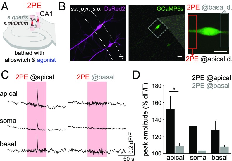
Two-photon pharmacology with alloswitch allows functional mapping of mGlu5 receptors at the subcellular scale in CA1 pyramidal neurons. (A) Experimental scheme in organotypic hippocampal slices: slices are bathed with 10 μM alloswitch and 50 μM DHPG (agonist), as well as 100 μM LY367385 to prevent mGlu1 activation. Silencing of mGlu5 by alloswitch is released locally, by targeting 2PE to the stratum oriens or stratum radiatum of CA1 (basal and apical dendritic regions of pyramidal neurons, respectively). (B) CA1 pyramidal neurons expressing the Ca2+ sensor GCaMP6s (Center and Right) and DsRed2 (Left), and zoomed view (Right) of the neuron imaged in C. The extent of 2PE is indicated by boxes (12 × 52 μm). (Scale bars, 20 μm.). (C) Calcium traces recorded in the somatic, apical and basal dendritic regions, while 2PE of alloswitch (red boxes, 780 nm, 1.5 Hz, 1 min) is confined to the apical (Left) or basal (Right) dendritic segments. Activation of mGlu5 receptors in the apical, but not basal, dendritic compartment induce broad Ca2+ signals in CA1 pyramidal neurons. (D) Quantification of Ca2+ responses as normalized peak amplitude, measured at the indicated cellular compartment during 2PE at the apical (black) or basal (gray) dendritic compartment. Paired t test, *P = 0.032; n = 4 cells from 3 animals.

