Fig. 4.
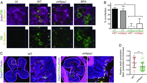
S. flexneri invasion disrupts cell polarity and colonic epithelium. (A) S. flexneri delocalizes the TfR to the apical domain of polarized Caco-2/TC7 cells. Cells were left uninfected (UI), infected with the WT or virAipaJ strains for 5 h, or treated with BFA for 30 min, before fixation without permeabilization and subsequent apical surface staining with an anti-TfR (OKT9) recognizing the extracellular domain of the receptor. (Scale bars: 10 µm.) (B) IpaJ and VirA enhance reinfection of polarized cells. Caco-2/TC7 cells were primo-infected with GFP-expressing WT or virAipaJ strains (green) for 4 h, and then reinfected with the same strains expressing dsRed for 2 more hours (red). The percentage of coinfection was quantified by determining the percentage of primo-infected cells (GFP positive) that were reinfected with dsRed-expressing bacteria. Mean ± SD. n = 4. ns, nonsignificant; *P < 0.05 (Kruskal–Wallis test followed by Dunn’s post hoc test). (C and D) S. flexneri WT has a deeper penetration than virAipaJ mutant into the guinea pig colonic tissue. (C) Colonic tissue from animals infected with GFP-expressing WT or virAipaJ mutant, 8 h postinfection, were stained with phalloidin-A647 (actin) and DAPI (nuclei). (D) The relative depth penetration of both strains into the colonic tissue was measured by their penetration from the epithelial surface compared with the total epithelial mucosa thickness at 4 h postinfection. Mean ± SD. 28 < n < 48 focus of infection pooled from two biological replicates. ***P < 0.001 (Mann–Whitney test).

