Fig. 1.
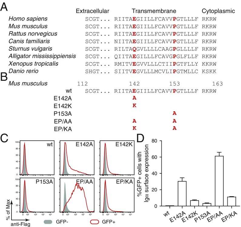
A conserved E-X10-P motif in the transmembrane region of Igα is responsible for its retention in ER. (A) Sequence comparison of Igα of different species. The conserved glutamic acid and proline are highlighted in red. (B) Sequence comparison of wt and mutant forms of Igα. The mutated amino acids are highlighted in red. (C) Flow cytometry analysis of the expression of Flag-tagged Igα on the surface of S2 cells transfected with plasmids encoding the indicated wt and mutant forms of Igα. Gray: GFP− untransfected cells; Red: GFP+ transfected cells. (D) Quantified Igα surface expression results presented as a bar graph. Data represent the mean and SE of a minimum of 3 independent experiments.

