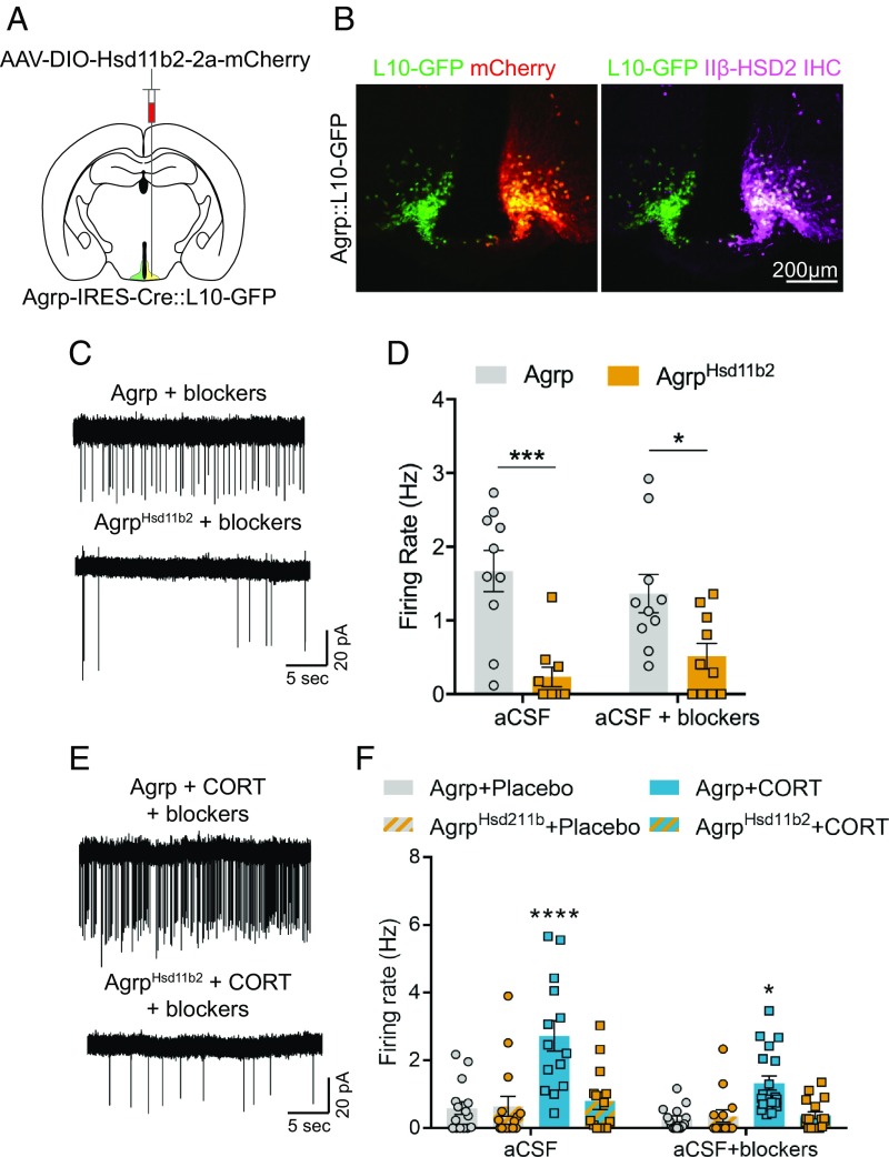
Fig. 5.
Expression of 11β-HSD2 in AgRP neurons prevents corticosterone-mediated increases in their firing rate. (A) Schematic of unilateral AAV-DIO-Hsd11b2-2a-mCherry injections into AgRP-Cre neurons in Agrp-IRES-Cre::L10-GFP mice. (B) Representative unilateral 11β-HSD2-mCherry expression (Left) and 11β-HSD2 immunohistochemical staining (Right) from an Agrp-IRES-Cre::L10-GFP mouse. (C) Representative cell-attached recordings of Agrp::L10-GFP (Top) and Agrp::L10-GFPHsd11b2 neurons (Bottom) from mice fasted for 16 h. (D) Summary of Agrp:L10-GFP and Agrp::L10-GFPHsd11b2 neuron firing rates from mice fasted for 16 h (Agrp-aCSF, n = 10 cells; Agrp-aCSF+blockers, n = 10 cells; AgrpHsd11b2-aCSF, n = 10 cells; AgrpHsd11b2-aCSF+blockers, n = 10 cells from 3 fasted and 3 fed mice). Two-tailed unpaired t tests with Holm–Sidak multiple-comparisons correction. *P < 0.05; ***P < 0.001. (E) Representative cell-attached recordings of Agrp::L10-GFP (Top) and Agrp::L10-GFPHsd11b2 neurons (Bottom) from mice implanted with corticosterone pellets. (F) Summary of Agrp::L10-GFP and AgRP::L10-GFPHsd11b2 neuron firing rates from mice implanted with placebo or corticosterone pellets (Placebo Agrp-aCSF, n = 15 cells; Placebo AgrpHsd11b2-aCSF, n = 15 cells; CORT Agrp-aCSF, n = 14 cells; CORT AgRPHsd11b2-aCSF, n = 14 cells; Placebo Agrp-aCSF+blockers, n = 15 cells; Placebo AgrpHsd11b2-aCSF+blockers, n = 14 cells; CORT Agrp-aCSF+blockers, n = 19 cells; CORT AgRPHsd11b2-aCSF+blockers, n = 17 cells from 3 placebo-implanted and 3 CORT-implanted mice). Two-way ANOVA with Tukey’s multiple-comparisons test. *P < 0.05; ****P < 0.0001. Data are the mean ± SEM.

