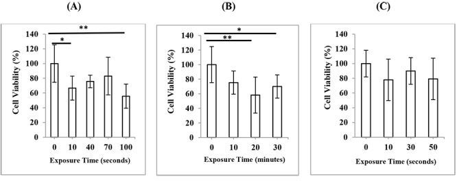
Figure2.

Cell viability of HDF cells, 24 h after exposure to ultrasound in (A) 3D mode (B) 4D mode (C) color Doppler mode of second-trimester fetal sonography condition. Data represent the mean ± SD of three independent experiments.* P < 0.05; ** P < 0.01.
