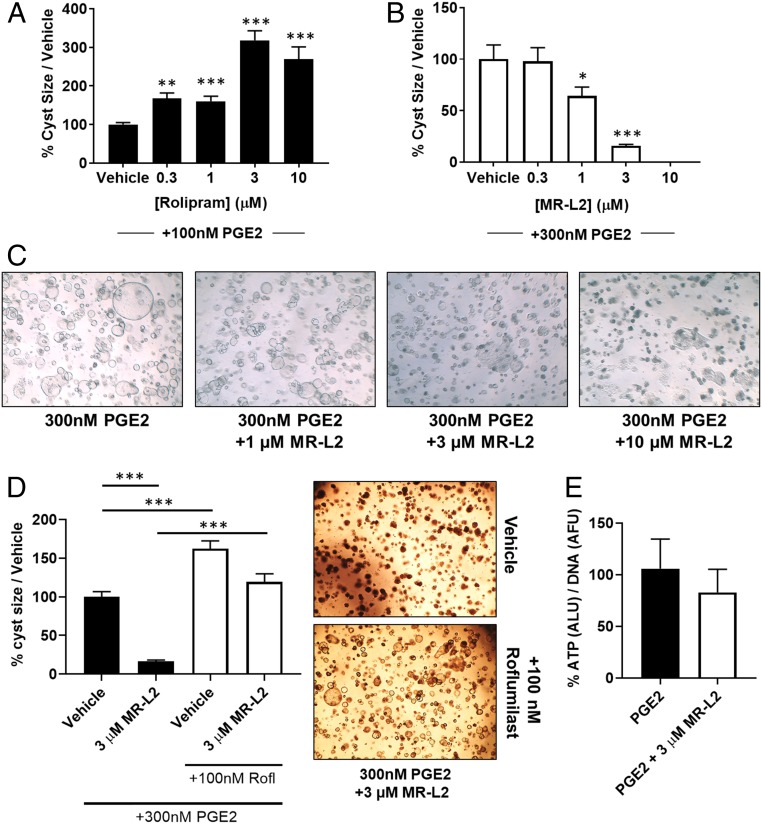
Fig. 4.
MDCK cyst formation is regulated by PDE4 activity and is suppressed by pharmacological activation of PDE4 long isoforms. (A) MDCK cells were grown in a 3D collagen matrix and treated with increasing concentrations of the PDE4-selective inhibitor, rolipram. Rolipram exacerbates the PGE2-induced formation of large cystic structures, demonstrating that PDE4 enzymes are localized within compartments that control cyst expansion. (B) PGE2-induced MDCK cell cyst formation was suppressed by MR-L2 (EC50, 1.2 µM). Data are displayed as a percentage of PGE2 stimulation alone, and error bars represent the SEM of all cysts measured in each condition. (C) Representative images of MDCK cysts treated with increasing concentrations of MR-L2. The images illustrate a concentration-dependent suppression of cyst size. (D) Cotreatment of PGE2 (300 nM)-stimulated MDCK cystic cultures with roflumilast (100 nM) and/or MR-L2 (3 µM) shows that inhibition of PDE4 catalytic activity ablates the suppressive effects of MR-L2 on cyst growth. Error bars represent the SEM. Representative images of MR-L2, and roflumilast/MR-L2–treated cultures are shown. (E) DNA normalized ATP concentrations within the cystic cultures were used as a measurement ATP in the cystic cultures. The balance of ATP signaling in the cystic cultures was not perturbed by MR-L2 treatment.

