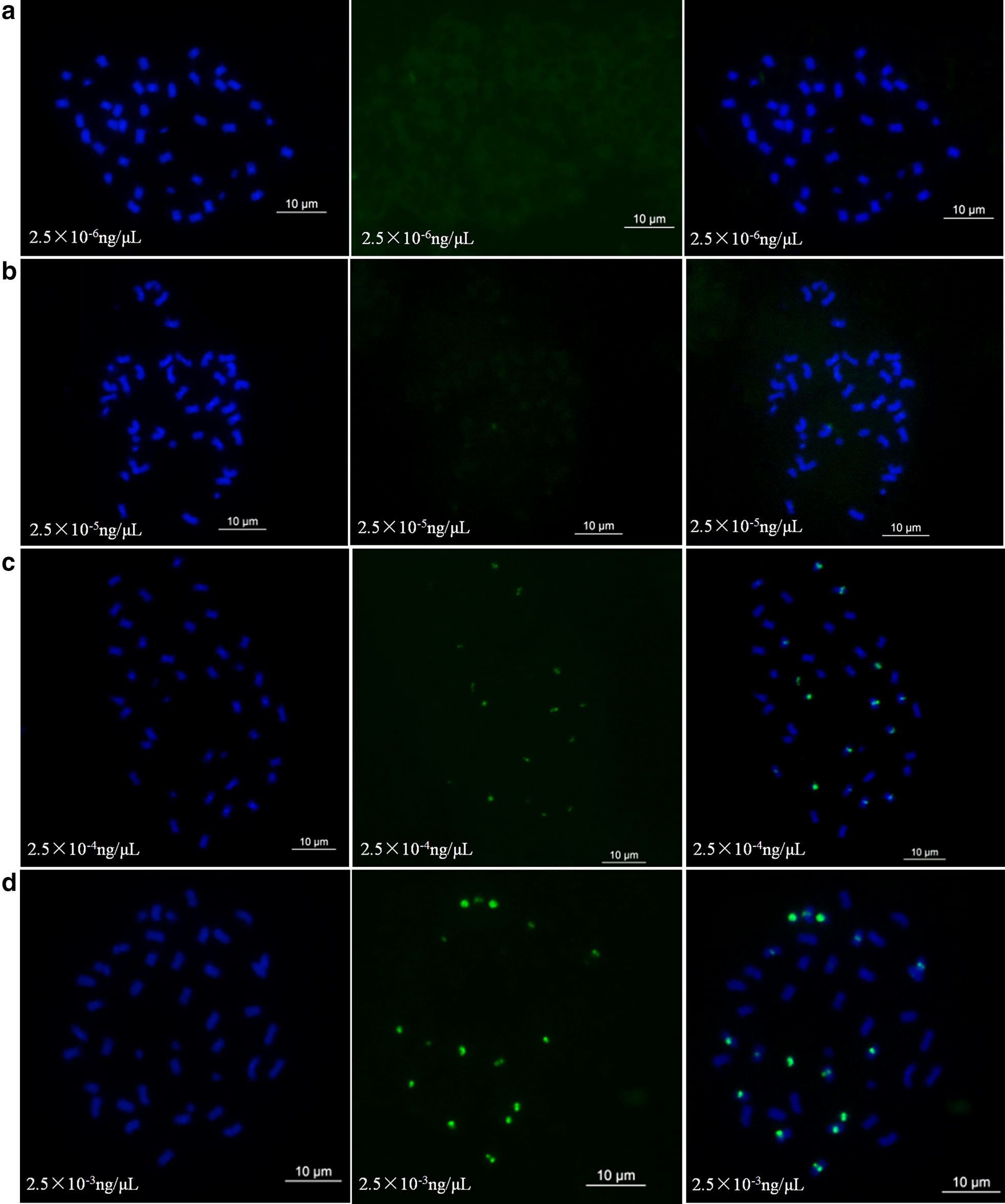
Fig. 1.

Concentration comparison analysis of oligonucleotide dye using DP-8 as a probe in Arachis hypogea cv. Silihong (SLH). Green signals show oligonucleotides that are DP-8 modified with FAM. a–d Show the results using oligonucleotide dye concentrations of 2.5 × 10⁻³ ng/µL, 2.5 × 10⁻⁴ ng/µL, 2.5 × 10⁻⁵ ng/µL, and 2.5 × 10⁻⁶ ng/µL, respectively
