
Figure 2.
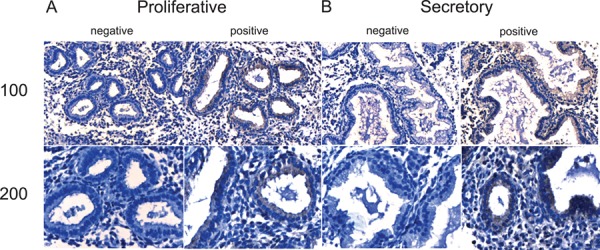
Immunohistochemical localization of MR1B in the endometrium across the menstrual cycle. Representative photomicrographs of MR1B expression at medium (×100) and high (×200) power magnification during the proliferative (A) and secretory (B) phases along with negative (without primary antibody) controls.