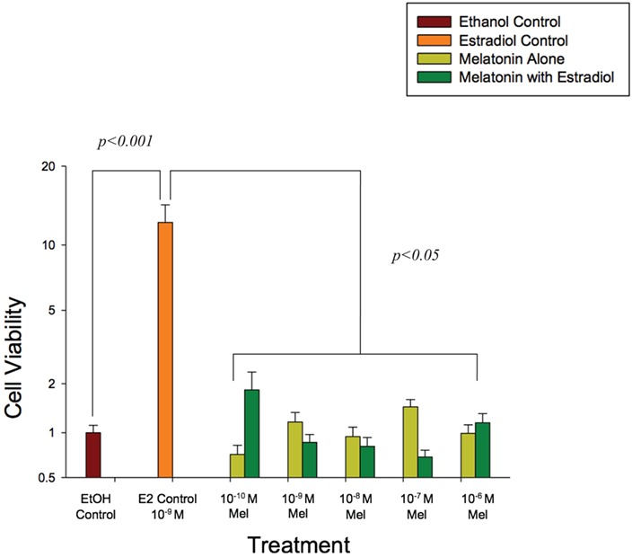
Figure 4.

Increasing concentrations of melatonin (0.1 nM–1.0 μM) alone and in the presence of estradiol (1.0 nM) attenuated endometrial epithelial cell proliferation at 48 hours of culture. Cell viability in the control wells was set to 1, and data are expressed on a log scale. Endometrial epithelial cells (CRL1671) were incubated in phenol red-free DMEM/F12 media supplemented with 10% charcoal-stripped FBS and 0.1% ITS. Results are the mean ± SEM of three separate experiments with eight replicates/concentration in each experiment. Data were analyzed by two-way ANOVA and Duncan’s multiple range test. A P < 0.05 was considered significant.
