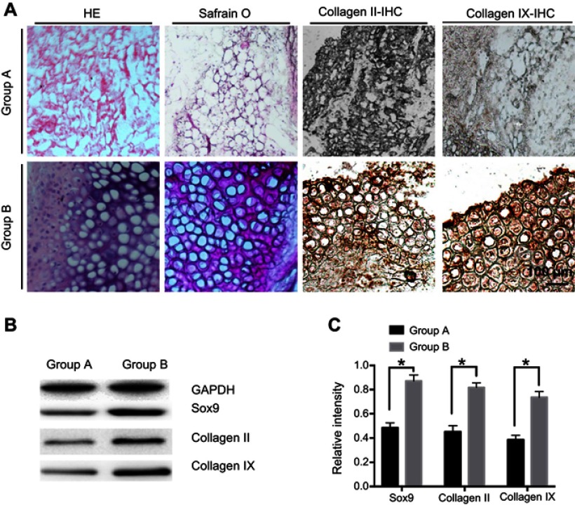
Figure 5.
In vivo chondrogenesis of rMSCs. (A) Immunohistochemical stain of different groups. (B) Western blot (WB) analysis of Sox9, collagen II, and collagen IX. (C) Densitometric analysis of Sox9, collagen II, and collagen IX. Group A, untransfected rMSCs with chitosan hydrogel; Group B, transfected rMSCs with chitosan hydrogel. *P<0.05.

