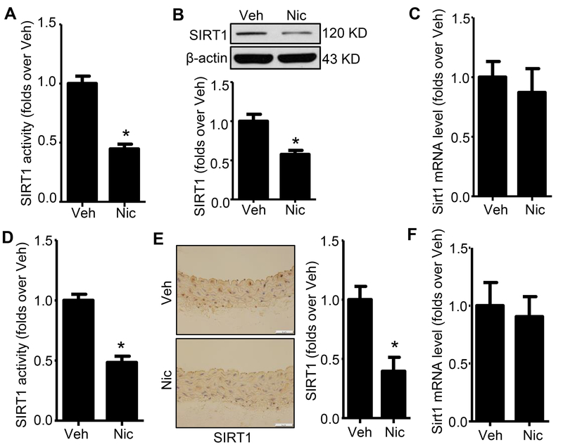
Figure 2. Nicotine reduces SIRT1 activity and protein expression in both cultured hASMCs and aorta.
A. SIRT1 activity was measured from hASMCs nuclear extracts. Data was normalized to the activity measured in vehicle-treated hASMCs (n=5; * P < 0.05 vs. Veh). B. Western blot analysis of SIRT1 protein expression in hASMCs treated with vehicle or nicotine for 24h (n=5; * P < 0.05 vs. Veh). C. Quantitative real-time PCR of Sirt1 in hASMCs treated with vehicle or nicotine for 24h (n=3). D. SIRT1 activity was measured from aorta nuclear extracts. Data were normalized to the activity measured in vehicle-treated mice (n=5; * P < 0.05 vs. Veh). E. Representative images of IHC staining and quantification of SIRT1 in the aorta from vehicle- and nicotine-treated mice (n=5; * P < 0.05 vs. Veh). F. Quantitative real-time PCR of Sirt1 in aorta from vehicle- and nicotine-treated mice (n=3). Veh, vehicle; Nic, nicotine. Negative control for anti-SIRT1 staining was presented in Online Figure II.

