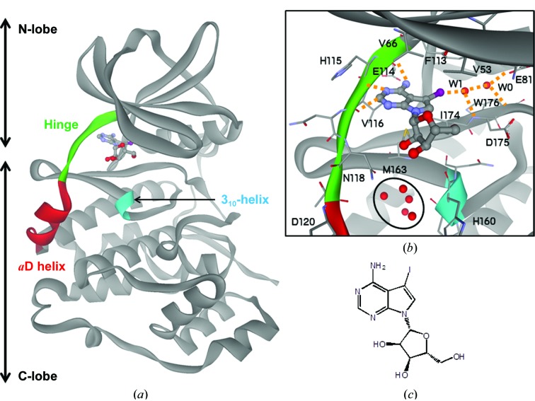
Figure 1.
Crystal structure of 5-iodotubercidin (5IOD) bound to the ATP-binding site of CK2a1. (a) Overall structure of the 5IOD–CK2a1 complex. (b) Mode of interaction of 5IOD with CK2a1. Hydrogen bonds are shown as orange dotted lines. The clustered water molecules in the αD pocket are shown as red spheres and are circled. (c) Chemical structure of 5IOD.

