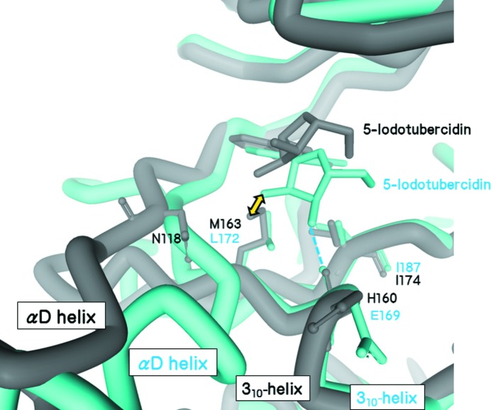
Figure 3.
Superimposition of the 5-iodotubercidin complexes of CK2a1 (grey) and CK1g2 (blue). The ribose moiety in the CK2a1 complex is located in the upper position when compared with that of CK1g2. The position of the ribose moiety in the CK1g2 structure would cause steric clashes with Met163 of CK2a1 (yellow arrow).

