1.
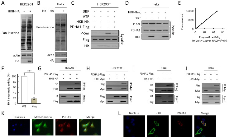
Hexokinase (HK) II interacts with and phosphorylates the alpha subunit of pyruvate dehydrogenase (PDHA1) in an ATP-dependent manner. (A) HA-tagged HKII was transfected into HEK293T cells and pan-phosphorylation level of serine (Pan-P-serine) was tested in HEK293T cell line; (B) HA-tagged HKII was transfected into HeLa cells and Pan-P-serine was tested in HeLa cell line; (C) Purified PDHA1 was treated with recombinant HKII in the presence and absence of ATP, and the levels of P-Ser of PDHA1 in the reaction mixture after treatment were determined; (D) Flag-tagged HKII was expressed in HeLa cells. Pan-P-serine levels of PDHA1 from cells cultured with and without 3BP (5 mmol/L) supplementation were determined; (E) Standard curve for HKII specific activity versus fluorescence (n=3); (F) Enzymatic activity of purified wild type HKII (WT) and HKIImut (Mut) were measured; (G) Flag-tagged PDHA1 and HA-tagged HKII were co-expressed in HEK293T cells. HKII co-purified with PDHA1 was detected by HA antibody; (H) Flag-tagged PDHA1 and Myc-tagged HKII were co-expressed in HEK293T cells. PDHA1 co-purified with HKII was detected by Flag antibody; (I) Flag-tagged PDHA1 and HA-tagged HKII were co-expressed in HeLa cells. HKII co-purified with PDHA1 was detected by HA antibody; (J) Flag-tagged PDHA1 and Myc-tagged HA were co-expressed in HeLa cells. PDHA1 co-purified with HKII was detected by Flag antibody; (K) PDHA1 was expressed in HeLa cells. PDHA1, mitochondria, and nucleus were marked with red, green, and blue fluorescence, respectively; (L) HKII and PDHA1 were co-expressed in HeLa cells. HKII, PDHA1, and nucleus were marked with green, red, and blue fluorescence, respectively. ***, P<0.001.

