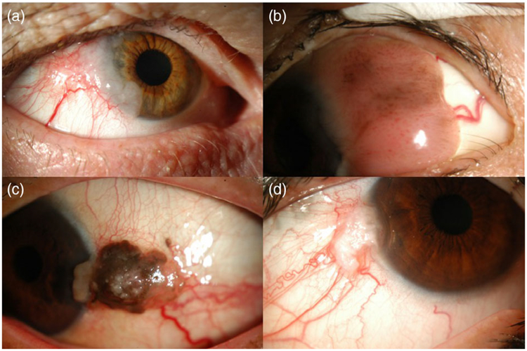
FIGURE 1.
Slit lamp photographs demonstrating a variety of clinical presentations of ocular surface squamous neoplasia. (a) Extensive gelatinous lesion on the temporal conjunctiva and extending onto the cornea from 7 to 11 o’clock, with large feeder vessels noted inferotemporally. (b) Large papillary mass on the temporal bulbar conjunctiva and extending onto the cornea. (c) Pigmented lesion involving the left temporal bulbar conjunctiva, with feeder vessels and extension onto the cornea. (d) Focal gelatinous white mass at the temporal limbus, with extension onto the cornea and with sentinel-type vessels

