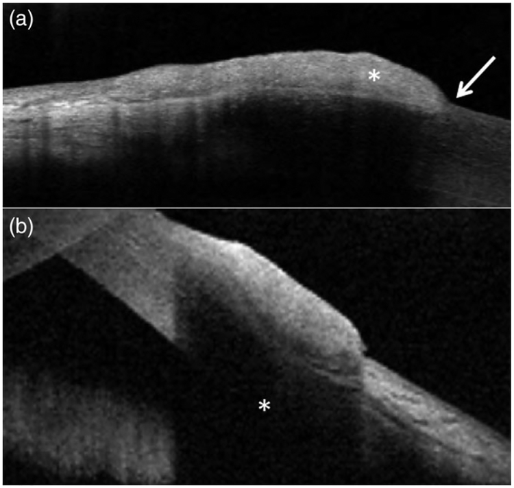
FIGURE 3.
Anterior segment optical coherence tomography (AS-OCT) images of ocular surface squamous neoplasia (OSSN). (a) AS-OCT image of a lesion demonstrating characteristic OSSN features, including a thickened, hyper-reflective epithelial layer (asterisk) and an abrupt transition between abnormal and normal epithelium (arrow); subsequently found to be invasive OSSN on biopsy. (b) AS-OCT image of a nodular OSSN lesion demonstrating substantial sub-epithelial shadowing (asterisk) underlying thickened, reflective epithelium; subsequently found to be non-invasive OSSN on biopsy. The degree of invasion beneath the epithelium in (a) and (b) could not be determined from AS-OCT images alone

