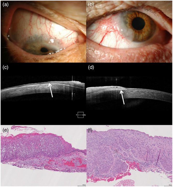
FIGURE 4.
Clinical, imaging and histopathologic appearances of non-invasive and invasive ocular surface squamous neoplasia (OSSN). (a) Slit lamp photograph of a superior gelatinous lesion with vascularity and extension onto the cornea. (b) Slit lamp photograph of a temporal gelatinous lesion with large inferotemporal vessels and extension onto the cornea. (c) Anterior segment optical coherence tomography (AS-OCT) image of the lesion from (a), demonstrating epithelial thickening and hyper-reflectivity consistent with OSSN as well as a lack of hyper-reflectivity at the epithelial base (arrow), possibly suggestive of more superficial epithelial disease. (d) AS-OCT image of the lesion from (b), demonstrating epithelial thickening and hyper-reflectivity as well as prominent sub-epithelial hyper-reflectivity (arrow), possibly suggestive of deeper invasion. (e) Histologic section of the lesion from (a) and (c), demonstrating moderate to focal severe epithelial dysplasia consistent with non-invasive OSSN. (f) Histologic section of the lesion from (b) and (d), demonstrating superficially invasive squamous cell carcinoma consistent with invasive OSSN

