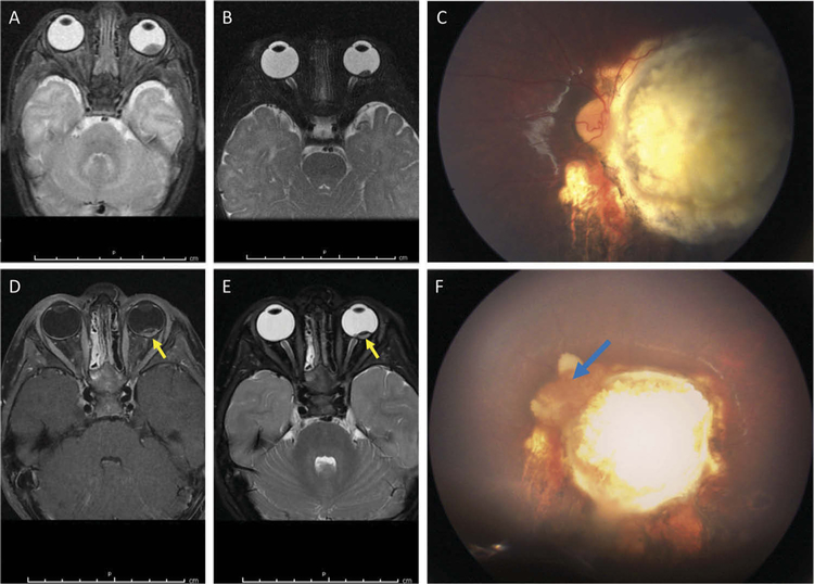
Figure 2.
Case 2: MRI and fundus photos prior to enucleation. (A) T2-weighted MRI image of the left intraocular tumor at diagnosis at 2 weeks of age. (B) T2-weighted image of the stable, calcified tumor at 8 months of age without abnormal enhancement. (C) At 35 months of age, there was a stable calcified tumor on funduscopic exam. (D, E) The T1-weighted (D) and T2-weighted (E) images 1 week prior to enucleation demonstrate increased enhancement (yellow arrows) at the posterior aspect. (F) Fundus image at the time of enucleation shows an active vascularized recurrence (blue arrow) at the nasal aspect of the calcified tumor, corresponding with the area of enhancement on MRI.

