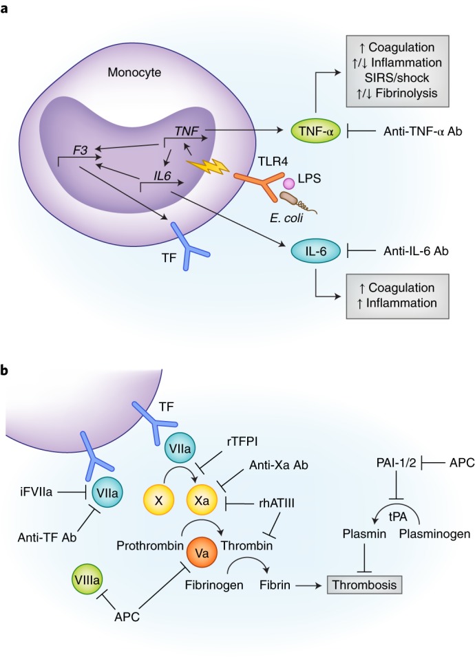
Fig. 1.

(a) Simplified rationale for performing studies in NHP sepsis models of antibody inhibition of TNF-α and IL-6. Lipopolysaccharide (LPS) or E. coli are shown binding to a TLR4 surface receptor of a monocyte which initiates downstream signal transduction (yellow lightning bolt) and transcriptional upregulation of TNF-α. TNF-α further activates other pro- and anti-inflammatory cytokines, including IL-6. Both cytokines promote hypercoagulability by upregulating tissue factor (TF) expression. TNF-α also simultaneously promotes and inhibits fibrinolysis via activation of tPA and PAI-1/2, respectively (see Fig. 1b). (b) Multiple therapeutics are shown inhibiting different components of the extrinsic coagulation pathway. These drugs were tested using LPS or E. coli NHP sepsis models. Abbreviations: Ab, antibody; APC, activated protein C; F3, factor 3; iFVIIa, site inactivated factor VIIa; IL6, interleukin 6; LPS, lipopolysaccharide; PAI-1/2, plasminogen activator inhibitor 1/2; rhATIII, recombinant human antithrombin III; SIRS, systemic inflammatory response syndrome; TF, tissue factor; TLR4, toll-like receptor 4; TNF, tumor necrosis factor.
