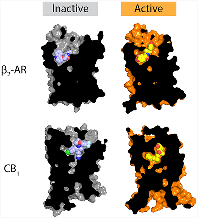
Figure 4.
Agonist recognition. The molecular details of agonist recognition are highly diverse, although most agonist-bound activate-state GPCR structures show a modest contraction of the ligand binding site relative to their inactive-state counterparts. Here, the structures of the inactive and active β2 adrenergic receptor (PDB entries 2RH1 and 4LDL, respectively) and the CB1 cannabinoid receptor (PDB entries 5U09 and 5XRA for the inactive and active states, respectively) are shown as representative examples, showing contraction of the binding site upon activation, as well as the far more extensive nature of structural rearrangements upon activation of the CB1 receptor compared to the β2 receptor.

