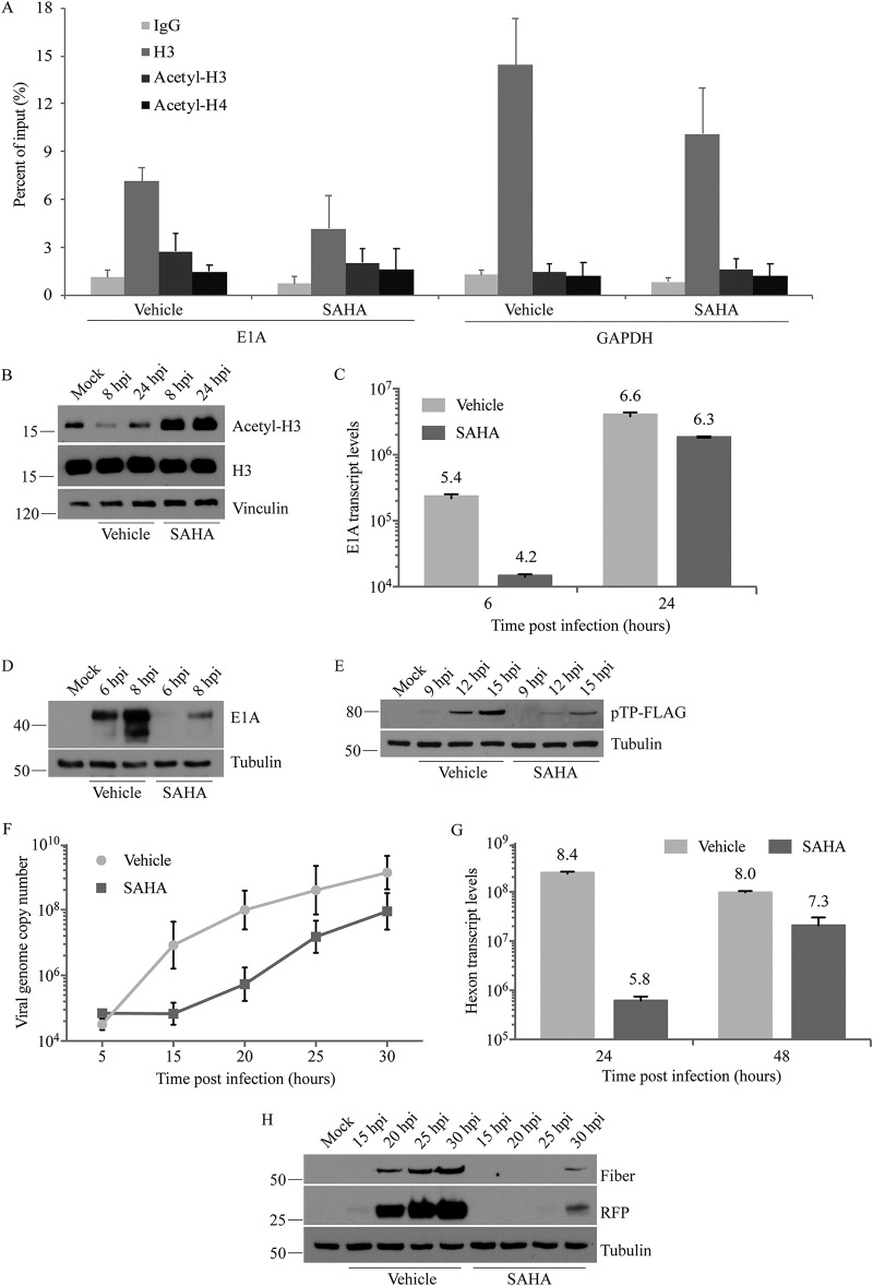
FIG 5.
SAHA impacts multiple stages of HAdV life cycle. A549 cells and 10 μM SAHA were used in all experiments. (A) Ad-late/RFP (MOI of 10) was used in a “cold infection” to synchronize virus entry into cells. The cells were treated with vehicle or SAHA and subjected to ChIP at 6 hpi with the indicated antibodies, followed by qPCR with primers specific to HAdV E1A or cellular GAPDH regions. (B) Lysates of cells infected with Ad-late/RFP (MOI of 10) and treated with SAHA were collected at 8 or 24 hpi for immunoblot analysis. (C and G) After infection and drug treatment as in panel B, total cellular RNA was extracted at the indicated times, and cDNA was generated by reverse transcription. qPCR analysis was conducted using primers to the HAdV E1A (C) or hexon (G) regions. (D and H) Infected, SAHA-treated cell lysates were collected at the indicated times for immunoblot analysis to detect viral early (D) and late (H) proteins. (E) Replication-competent Ad(E1+)TP-F was used at an MOI of 50 for infection to assess expression from the E2 region in the presence and absence of SAHA. (F) Infection and drug treatment were carried out as in panel B, and genomic DNA was extracted at the indicated times for qPCR using primers specific to hexon. SAHA did not affect HAdV entry, DNA association with H3, or acetyl-H3/acetyl-H4 levels, but it inhibited the expression of early and late genes and viral DNA replication. All error bars represent the SD (n = 4 for ChIP and n = 2 for all other experiments).

