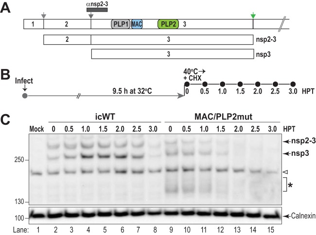
FIG 6.
Mutations in the macrodomain and PLP2 alter the stability of replicase protein nsp3. HeLa-MHVR cells were infected with either icMHV-WT or MAC/PLP2mut virus (MOI of 5) and incubated at 32°C for 9.5 h. Then 20 μg/ml of CHX was added, and the cells were shifted to the nonpermissive temperature. Lysates were prepared every 30 min, the proteins were separated by SDS-PAGE, and nonstructural proteins nsp2-3 and nsp3 were visualized by immunoblotting. (A) Schematic diagram of MHV replicase polyprotein indicating the processing pathway and the region identified by the anti-nsp2-3 antibody. (B) Outline of the experiment. (C) Western blot evaluating the level of nsp2-3 and nsp3 proteins detected after a shift to the nonpermissive temperature. These are representative data from two independent experiments. The arrowhead indicates detections of cellular protein in all lysates. The asterisk indicates degradation products detected by anti-nsp2-3 antibody in the MAC/PLP2mut virus-infected cells.

