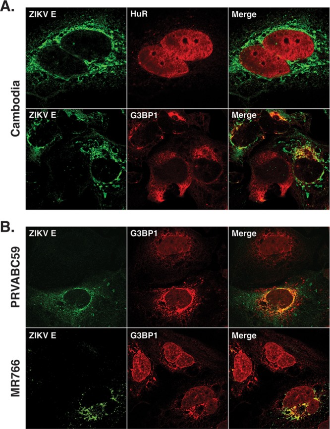
FIG 6.

G3BP1 colocalizes with ZIKV E protein. One day postinfection, Huh7 cells infected with ZIKV at an MOI of 5 were fixed, permeabilized, and prepared for confocal imaging. (A) Localization of ZIKV E protein with HuR and G3BP1 following infection with the Cambodia strain of ZIKV. (B) Localization of ZIKV E protein with G3BP1 following infection with the Puerto Rican (PRVABC59) and Ugandan (MR766) strains of ZIKV. The immunofluorescence images shown are representative of at least three independent experiments.
