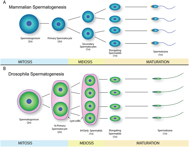
Fig. 3.
Mammalian and Drosophila spermatogenesis. (A) Schematic representation of mammalian spermatogenesis from spermatogonium to spermatozoa, depicting mitosis, meiosis, and maturation stages. (B) Drosophila spermatogenesis schematic representation, where spermatozoa have a more elongated nucleus and lack of acrosome region when compared with mammalian spermatozoa. 2n, diploid cells; 1n, haploid cells.

