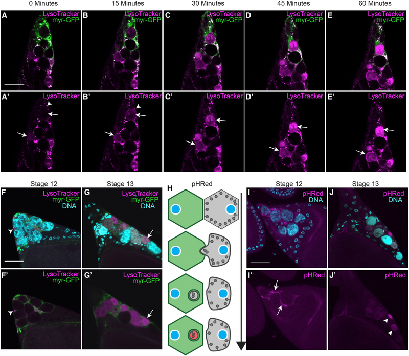
Figure 1.
Nurse Cells Are Surrounded by Stretch Follicle Cells and Acidified (A–E) Time lapse images of stretch follicle cell (SFC) > myr-GFP (green) stage 13 egg chamber labeled with LysoTracker (LT, magenta).
(A’–E’) The same images with the LT channel only. LT puncta accumulate around nurse cells (NCs) within SFCs (arrowheads in A’ and B’) as NCs become acidified (arrows in A’–E’) over 60 min.
(F and G) SFC>myr-GFP stage 12 (F) and stage 13 (G) egg chambers stained with DAPI (cyan) and LT (magenta).
(F’ and G’) The same egg chambers showing only the GFP and LT channels.
(F and F’) LT puncta accumulate around NCs in stage 12 (arrowhead).
(G and G’) NCs are acidified in stage 13 (arrow).
(H) Diagram of pHRed as an acidification detector, adapted from Fishilevich et al., (2010). pHRed is targeted to the cytoplasmic side of the plasma membrane and fluoresces red upon acidification.
(I and J) Germline > pHRed egg chambers stained with DAPI (cyan).
(I) Acidification of NC membrane detected by pHRed in stage 12.
(J) NCs in stage 13 are pHRed positive. Scale bars, 50 μm.

