
Figure 5.
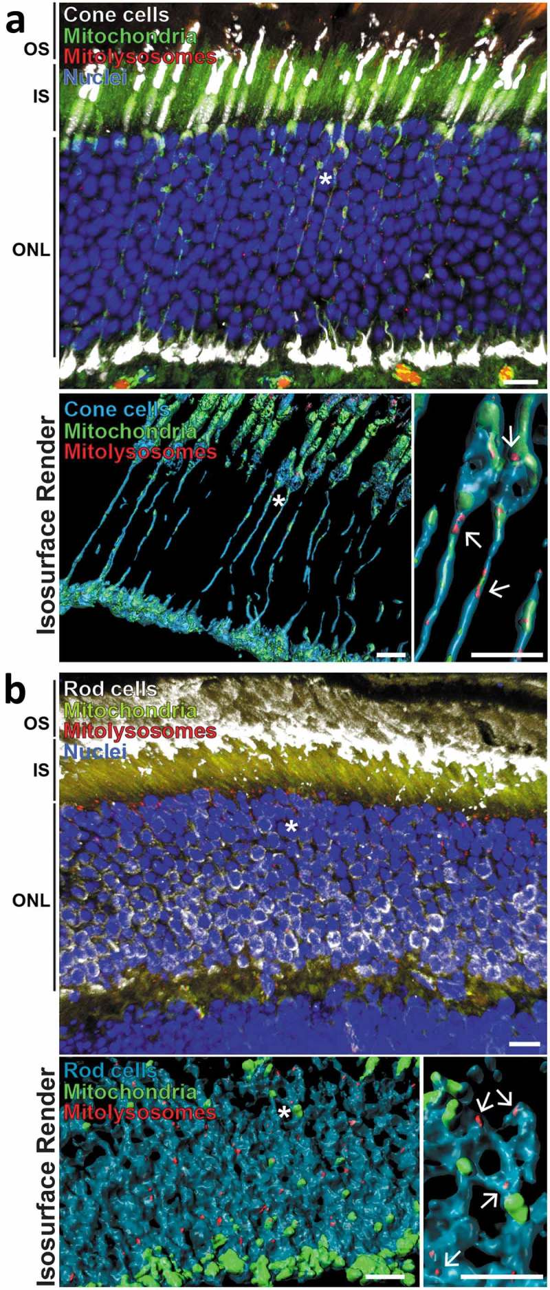
Mitophagy occurs in retinal photoreceptor neurons. (a) Z-projection showing adult mito-QC retinal outer nuclear layer (ONL) stained with antibodies against ARR3/cone arrestin (white, top panel). Inner segments (IS) and outer segments (OS) of the photoreceptor cells are also indicated. Lower left panel shows isosurface render of cone cells (cyan) from the above micrograph. Asterisk denotes cone projections magnified in lower right panel, arrows indicate mitolysosomes. Note presence of mitolysosomes in cone processes. (b) Z-projection of mito-QC retinal outer nuclear layer (ONL) and inner and outer photoreceptor cell segments (IS and OS respectively) stained with antibodies against SAG/visual arrestin to identify rod cells (white, top panel). Lower left panel shows isosurface render of rod somata (cyan) from the above micrograph. Asterisk denotes area of cells shown magnified in lower right panel and arrows mark mitolysosomes. Scale bars: 10 μm.