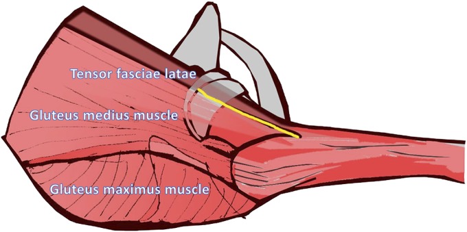
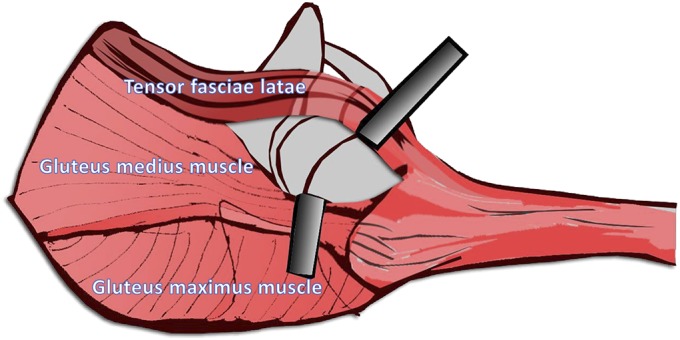
Figs. 3-A and 3-B The anterior border of the gluteus medius muscle and the posterior border of the tensor fasciae latae were identified and separated from each other along the yellow line.
Fig. 3-A.

Fig. 3-B.
The gluteus medius muscle was retracted posteriorly, and the tensor fasciae latae was retracted anteriorly to allow entry into the hip joint.

