Figure 2.

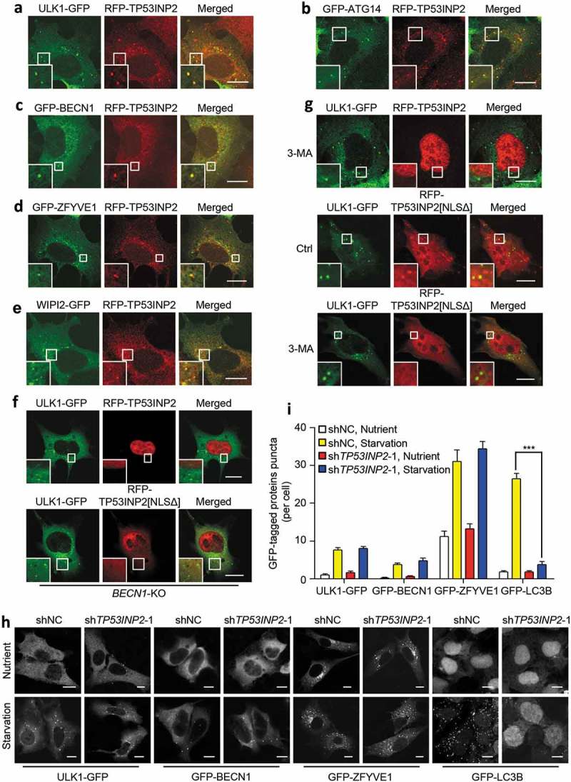
Association of TP53INP2 with early autophagic structures. (a-e) Colocalization of RFP-TP53INP2 with ULK1-GFP (a), GFP-ATG14 (b), GFP-BECN1 (c), GFP-ZFYVE1 (d) or WIPI2-GFP (e) in starved MEFs. (f and g) Localization of RFP-TP53INP2 or RFP-TP53INP2[NLSΔ] in starved BECN1-KO (f) or 3-MA-treated (g) HEK293 cells. The cells were imaged by confocal microscope 24 h after cotransfection of the plasmids. (h) Formation of puncta in HEK293 cells stably expressing ULK1-GFP, GFP-BECN1, GFP-ZFYVE1 or GFP-LC3B. The cells were infected with lentivirus expressing non-silencing control shRNA or TP53INP2 shRNA for 72 h, with or without cell starvation for 2 h. (I) Quantification of the puncta in (h). The data are presented as mean ± SEM, n = 30 cells. ***, P < 0.001. Scale bars: 10 µm.
