Figure 3.

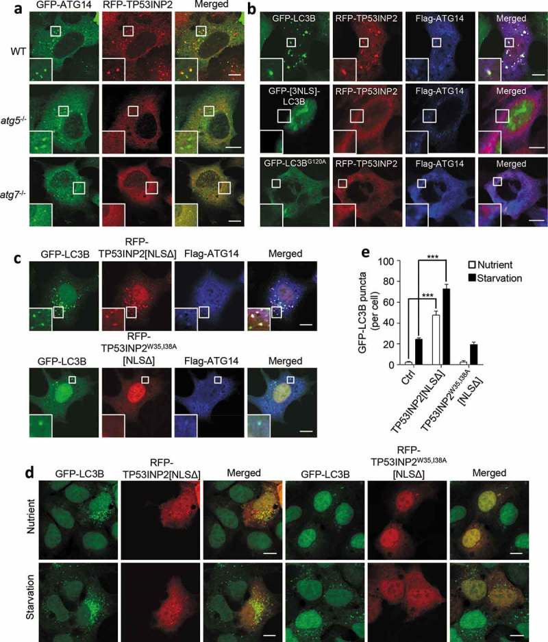
LC3 mediates the association of TP53INP2 with autophagic membranes. (a) Colocalization analysis of RFP-TP53INP2 and GFP-ATG14 in starved WT, atg5−/- or atg7−/- MEFs. (b) Colocalization analysis of RFP-TP53INP2 and Flag-ATG14 in starved HEK293 cells coexpressing GFP-LC3B, GFP-[3NLS]-LC3B or GFP-LC3BG120A. (c) HEK293 cells stably expressing GFP-LC3B were cotransfected with Flag-ATG14 and RFP-TP53INP2[NLSΔ], or with Flag-ATG14 and RFP-TP53INP2W35,I38A[NLSΔ]. Cells were then immunostained with anti-Flag and imaged with confocal microscopy. (d) GFP-LC3B punctum formation in HEK293 cells stably expressing GFP-LC3B with or without cell starvation. The cells were transiently transfected with RFP-TP53INP2[NLSΔ] or RFP-TP53INP2W35,I38A[NLSΔ]. (e) Quantification of GFP-LC3B puncta in (d). The data are presented as mean ± SEM, n = 30 cells. ***, P < 0.001. Scale bars: 10 µm.
