Figure 5.

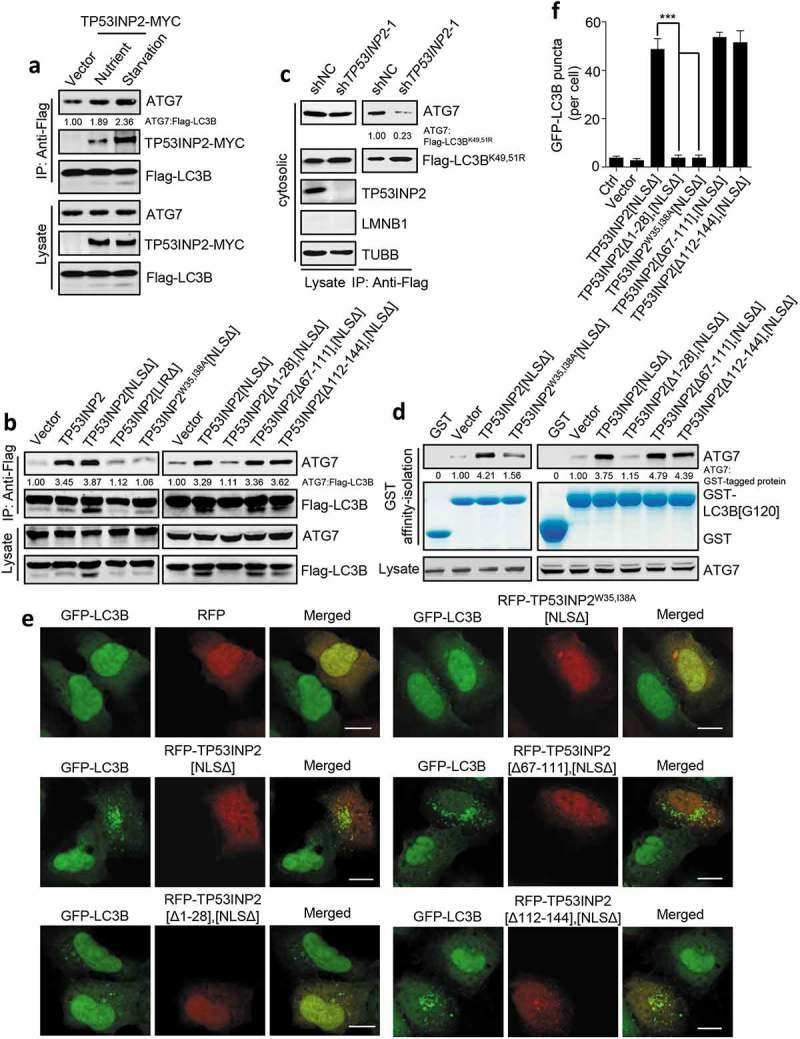
TP53INP2 facilitates LC3B-ATG7 interaction. (a) Coprecipitation of endogenous ATG7 with exogenous Flag-LC3B in TP53INP2-MYC cotransfected HEK293 cells with or without cell starvation. Flag-LC3B was immunoprecipitated using anti-Flag, then ATG7 and TP53INP2-MYC were detected by anti-ATG7 and anti-MYC respectively. (b) Coprecipitation of ATG7 with Flag-LC3B from HEK293 cells transiently expressing RFP-tagged TP53INP2 or each of the indicated TP53INP2 mutants. Flag-LC3B was immunoprecipitated using anti-Flag. (c) HEK293 cells stably expressing non-silencing shRNA or TP53INP2 shRNA were transfected with Flag-LC3BK49,51R and starved. The cells were then fractionated by differential centrifugation. Flag-LC3BK49,51R was immunoprecipitated from the cell cytosol using anti-Flag and the coprecipitated ATG7 was detected by western blot. (d) In vitro affinity-isolation assay of LC3B[G120]-ATG7 interaction. Purified GST-LC3B[G120] was incubated with cell lysate from HEK293 cells expressing the indicated RFP-tagged TP53INP2 mutants. After affinity-isolating GST-LC3B[G120] using glutathione-sepharose 4B beads, GST-LC3B[G120]-bound ATG7 was analyzed by western blot. (e) Confocal images of HEK293 cells stably expressing GFP-LC3B and transfected with plasmids expressing each of the indicated RFP-tagged TP53INP2 truncated mutants. (f) Quantification of GFP-LC3B puncta in (e). The data are presented as mean ± SEM, n = 30 cells. ***, P < 0.001. Scale bars: 10 µm.
