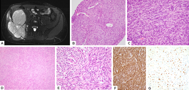
Figure 1: MEIS1-NCOA2 positive iliac bone rhabdomyosarcoma.
(case 1, 22/M). A) MRI showing a large tumor centered in the iliac bone with extraosseous extension. B) Pre-therapy biopsy shows a highly cellular spindle cell neoplasm with scattered branching vessels and insignificant stromal component; C) higher power with monomorphic spindle cells arranged in a streaming pattern. D, E) Resected tumor s/p chemotherapy reveals spindle cells in intersecting fascicles and scant eosinophilic cytoplasm. Immunohistochemical stains show F) diffuse positivity for desmin and G) focal Myogenin staining.

