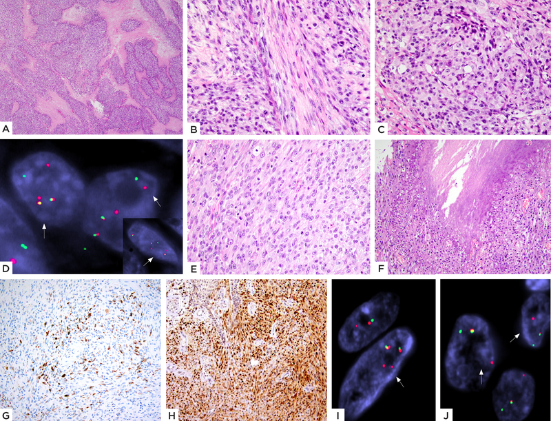
Figure 4: EWSR1-TFCP2 fusion positive rhabdomyosarcoma with a mixed spindle and epithelioid histology.
(A-D) Case 3 (27/F, skull): A) Low power showing a cellular neoplasm with geographic areas of necrosis. High power showing B) spindle cells with pale eosinophilic cytoplasm arranged in intersecting fascicles at 90-degrees and C) areas with distinct epithelioid features. D) FISH studies show break-apart signals for both TFCP2 and EWSR1 (inset) genes (red, centromeric, green, telomeric). (E-J) Case 4 (33/F, maxilla): E) Cellular neoplasm composed of plump ovoid to short spindle cells with moderately abundant pale eosinophilic cytoplasm, arranged in solid sheets or streaming pattern and F) areas of necrosis. Immunohistochemical stains show G) patchy positivity for desmin and H) relatively diffuse nuclear staining for MyoD1. FISH analysis showed the presence of break-apart split signals for both I) EWSR1 and J) TFCP2 genes (red, centromeric; green, telomeric)

