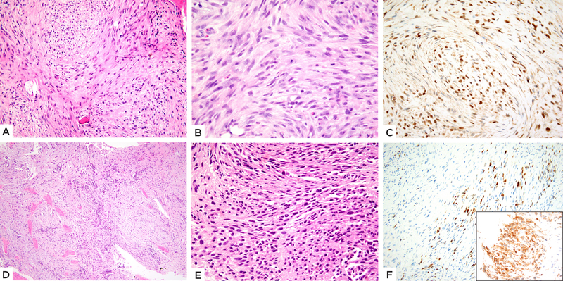
Figure 5: TFCP2 fusion positive subset showing mainly spindle cell morphology.
(A-C) EWSR1-TFCP2 fusion positive femur rhabdomyosarcoma (case 5, 20/M). A) Cellular neoplasm involving the bone and completely replacing marrow spaces with spindle cells arranged in intersecting fascicles or vague storiform, with B) pale eosinophilic cytoplasm and ovoid to fusiform nuclei with high mitotic activity. C) Immunohistochemical stain shows diffuse MYOD1 positivity. (D-F) FUS-TFCP2 positive iliac bone tumor (case 6, 37/F). D) Low power showing a cellular neoplasm diffusely involving bone. E) High power reveals short spindle cells arranged in short fascicles. F) Immunohistochemical stain shows focal Myogenin positivity (Inset: diffuse ALK immunoreactivity).

