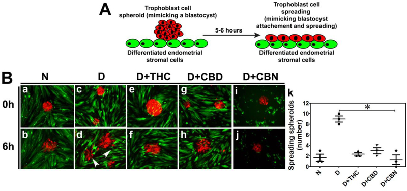
Figure 4. THC, CBD and CBN compromise trophoblast-endometrium interaction.
A. A schematic representation of the spheroid attachment assay. TB spheroids made from BeWo cells expressing mCherry that mimic trophoectoderm of the blastocyst were placed on a monolayer of undecidualized control or decidualized THESCs with or without THC, CBD or CBN treatment. Spheroid attachment and expansion were then assessed visually after 6 h of incubation. B. Shown are fluorescence images. TB spheroids prepared from mCherry-expressing BeWo cells (R-BeWo shown in red) were placed onto a monolayer of undecidualized control or decidualized EGFP-expressing THSECs (G-THSECs shown in green). Images a and b show that R-BeWo spheroids placed on undecidualized G-THSECs weakly attached and TB cells never migrated from the spheroids after 6 h. Images c and d show that R-BeWo spheroids placed on decidualized THESCs strongly attached and TB cells started migrating away from the spheroids after 6 h. Images e-j show that R-BeWo spheroids placed on decidualized THESCs in the presence of 0.5 μM THC (e and f), CBD (g and h) or CBN (i and j) had decreased attachment and delayed expansion after 6 h. N and D in panel B represent undecidualized and decidualized THESCs, respectively. k shows a significant decrease in the number of spreading R-BeWo spheroids placed on G-THESC decidualized in the presence of THC, CBD or CBN compared to control decidualized THESCs with no CB treatments. *indicates a p value of ≤ 0.05 between with and without CB treatments of decidualized THESCs.

