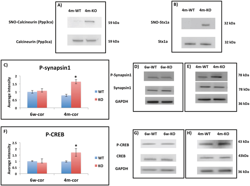
Fig. 3.

a Representative WB from eluted SNO-proteins prepared from 4m-WT and 4m-KO showing SNO-CN (Ppp3ca) in KO and not present in WT. CN shows similar levels in both groups. b Representative WB from eluted SNO-proteins prepared from 4m-WT and 4m-KO showing SNO-Stx1a in KO and not present in WT. Stx1a shows similar levels in both groups. c The relative average WB intensity of P-synapsin1 (Ser 62, Ser 67) comparing 6w-cor-WT to 6w-cor-KO and 4m-cor-WT to 4m-cor-KO. The data shows significant increase of P-synapsin1 in 4m-cor-KO compared to 4m-cor-WT. The data is normalized to synapsin1 and GAPDH and presented as mean ± SEM. One tailed t-test was conducted. *P < 0.05. WT mice (n = 4) and KO mice (n = 4). d Representative WB bands of P-synapin1, synapsin1, and GAPDH from cortex tissue prepared from 6w-WT and 6w-KO mice groups. e Representative WB bands of P-synapin1, synapsin1, and GAPDH from cortex tissue prepared from 4m-WT and 4m-KO. f The relative average WB intensity of P-CREB (Ser133) comparing 6w-cor-WT to 6w-cor-KO and 4m-cor-WT to 4m-cor-KO. The data shows significant increase of P-CREB in 4m-cor-KO compared to 4m-cor-WT. The data is normalized to CREB and GAPDH and presented as mean ± SEM. One tailed t-test was conducted. *P < 0.05. WT mice (n = 4) and KO mice (n = 4). g Representative WB bands P-synapin1, synapsin1, and GAPDH from cortex tissue prepared from 6w-cor and 6w-KO. h Representative WB from cortex tissue prepared from 4m-WT and 4m-KO
