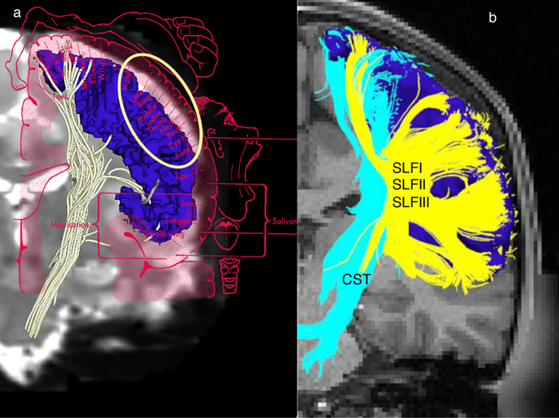
Fig. 4.
a A representative corticospinal tract (CST) of a healthy human subject delineated by diffusion magnetic resonance imaging (dMRI) tractography using high-resolution data of the Human Connectome Project (HCP) (Washington University; abbreviated as WashU) sample. Please note the paucity of fibers in the lateral aspect of the hemispheres. This is possibly due to limitations of dMRI tractography.
When overlaid with the motor homunculus (coronal plane), the lateral part of the motor homunculus indicates that the hand motor area, which should have been a part of the CST, is significantly limited, something that can possibly be explained by the presence of massive superior longitudinal fascicles I, II and III (SLF I, II and III) crossing fibers at the centrum ovale.
b In this representative case, the entire precentral cortex (dark blue) has been used as the seed to grow the CST (light blue).
Crossing fibers at the centrum ovale relating principally to the SLF I, II and III (in yellow) (Makris et al. 2005) do not allow effective sampling of CST from the lateral aspect of the precentral gyrus (motor cortex).

