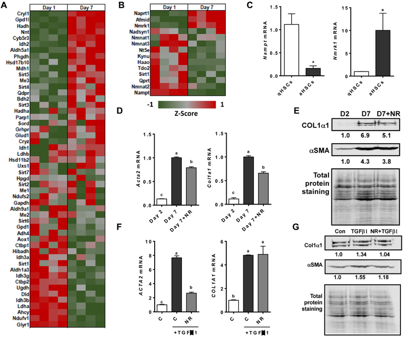
Figure 5.
NAD+ metabolism is altered in activated HSCs and NR treatment can attenuate the activation of hepatic stellate cells. A) Heat map of genes from RNA-Seq requiring NAD+ as a cofactor in quiescent HSCs (day 1) and activated HSCs (day 7). B) Heat map of genes from RNA-Seq of NAD+ de novo biosynthesis and salvage pathway in quiescent HSCs (day 1) and activated HSCs (day 7). C) mRNA expression of Nmrk1 and Nampt1 in mouse primary HSCs at day 1 and day 7. D) mRNA expression of primary mouse HSCs treated with or without 1.0 mM NR from day 2 to day 7. E) Representative Western blot analysis of mouse primary HSCs treated with or without 1.0 mM NR from day 2 to day 7 and normalized with total protein staining. The result of quantification is shown under each protein band. F) mRNA expression and G) Western blot analysis of primary human HSCs pretreated with 1.0 mM NR for 6 hours and then treated with 2 ng/mL of TGFβ1 for 24 h. The result of quantification is shown under each band. Data shown are mean ± SEM. Bars with different letters are significantly different from each other. (p < 0.05)

