. 2019 Jun 26;28(3):390–403. doi: 10.5607/en.2019.28.3.390

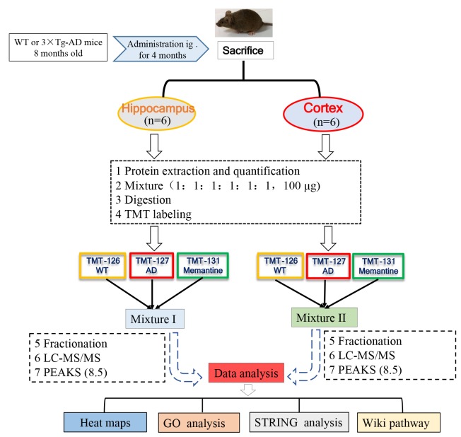
Fig. 1. Schematic diagram of experimental design. 3xTg-AD (AD) mice were treated with memantine (5 mg/kg, bid, ig) or equal volume of saline. The mice were sacrificed after behavioral assessment, and brain tissues were isolated for further proteomic analysis. Protein was pooled from six animals in each group (WT, AD or Memantine group) and was digested into peptides. The peptides were further labeled with 6-plex TMT and HPRP fractionation followed by LC-MS/MS analysis. Proteomics analysis was performed by using PEAKS 8.5 software, and bioinformatics analysis (Heat map, GO, STRING and Wiki path) was performed using DAVID 6.8 and Cytoscape software.

Fig. 1