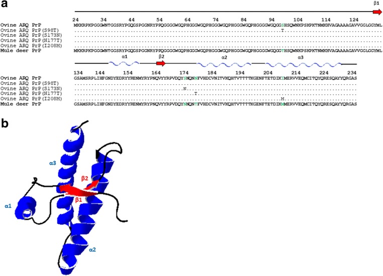
Fig. 3.
a Sequence alignment of the C-terminal domain (residues 134–234) of ovine PrP compared with mule deer PrP. Note the four amino acid differences that where substituted giving rise to four mutants: ovine ARQ PrP (S98T), ovine ARQ PrP (S173N), ovine ARQ PrP (N177T), and ovine ARQ PrP (I208M). b Diagram of ovine PrP, and the locations of the native secondary structures in sheep ARQ PrP (134–234) are indicated: the α-helical regions are represented in blue and the β-sheet region in red. The diagram was generated using the program Swiss-PdbViewer (4.1.0)

