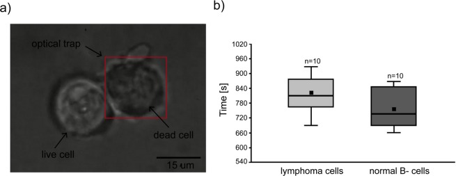
Figure 3.
(a) Trypan blue accumulation on the surface of untreated DLBCL cell (left), while the dead cell was held in optical trap > 800 s at 100 mW of laser power what resulted in cell membrane rupture and entering the dye inside the cell. The red frame indicates the area of operating range of the optical trap, while the focused laser beam is located in the centre of the trapped cell. The scalebar is set at 15 μm. (b) Comparison of the normal and lymphoma primary cells viability exposed to laser power of 100 mW using Trypan blue. Manipulation over 756 ± 73.24 s and 819 ± 72.31s for normal and lymphoma cells, respectively damaged the cell membrane.

