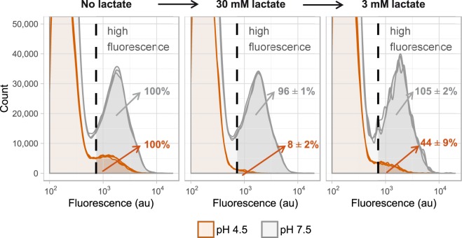
Figure 5.
Metabolic activity of ampicillin treated L. lactis MG1363. Stationary L. lactis MG1363_GFP cells were diluted in medium with ampicillin (n = 3). After 48 hours (left panel) cells were temporarily exposed to high lactate levels (middle panel). At pH 4.5 this reduced the GFP fluorescence due to weak acid uncoupling. When the lactate concentration was lowered the amount of highly fluorescent cells increased from 8 ± 2% to 44 ± 9% (right panel). This shows that 36 ± 7% of the cells could restore the high fluorescence signal, indicating that these cells were metabolically active.

