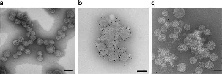
Fig. 1.
Electron microscopy of OMPC and Pfs230-OMPC conjugate. a Transmission electron microscopic image of OMPC. b Transmission electron microscopic image of Pfs230-OMPC-3 stored at 4 °C for one year, after incubation with anti-Pfs230 antibody, 4F12 (1:10), followed by gold-labeled secondary antibody. c Transmission electron microscopic image of Pfs230-OMPC-3 incubated with gold-labeled secondary alone (omitting incubation with primary antibody, 4F12). Images were taken on a FEI Biotwin Tecnai microscope and collected on an AMT XR611 camera system. Scale bar: 100 nm

