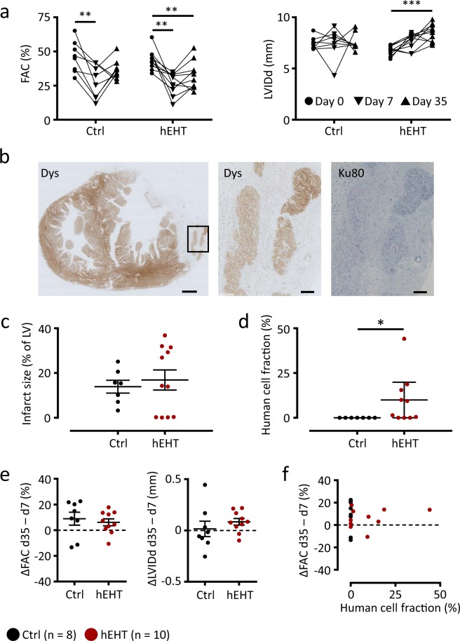
Figure 2.
Functional parameters, infarct size and human cell fraction. (a) Fractional area change (FAC) and left-ventricular inner diastolic diameter (LVIDd) in the control and hEHT group immediately before cryo-injury (day 0, ●), before graft implantation (day 7 after injury, ▼) and at the end of the observation period (day 35 after injury, ▲). (b) Representative transversal section at low magnification (left panel) and high magnification of the area indicated by the box (middle and right panel). Sections were stained for dystrophin (Dys) and Ku80 to validate human origin. Scale bars represent 1 mm (left panel) and 200 µm (middle and right panel). (c) Infarct size (% of left ventricle) and (d) mean human cell fraction (%) determined within the grafts. (e) Effect of graft implantation on fractional area change (FAC) and left-ventricular inner diastolic diameter (LVIDd). Values were calculated as differences between day 7 and day 35 following cryo-injury (ΔFAC and ΔLVIDd). (f) Correlation analysis between ΔFAC (ΔLVIDd) and human cell fraction. Data are given (a) as before-after plots and as individual data points with lines and error bars denoting (c) mean ± SEM and (d) median ± interquartile range, respectively. (e) Lines and error bars denote mean ± SEM. Results were analyzed with (a) an one-way ANOVA followed by multiple comparisons using a Tukey test with multiplicity adjusted P values and with (c,e) a parametric and (d) with an unparametric (Mann-Whitney) unpaired Student’s t-test. *P < 0.05, **P < 0.01, ***P < 0.001. Ctrl, animals implanted with cell-free fibrin constructs (n = 8); hEHT, animals with implanted hEHTs (n = 10).

