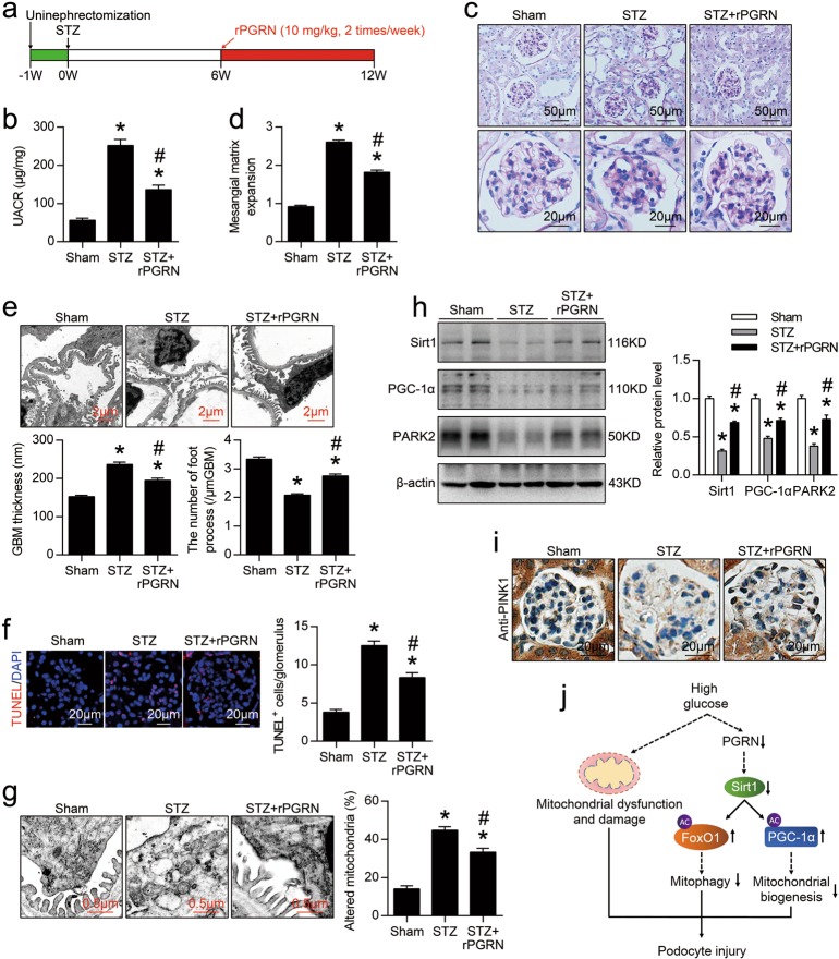
Fig. 8. Administration of recombinant human PGRN (rPGRN) protected against podocyte injury in mice with DN.
a A representative figure showing the procedure of rPGRN treatment in this study. b Urine albumin-to-creatinine ratio (UACR) in different groups of mice. c Representative images of PAS staining of kidney sections in different groups of mice. d Quantification of Mesangial Matrix Index of glomerulus from different groups of mice. e Representative TEM images showing morphological changes in the podocyte foot process in different groups of mice. Quantitative assessment of glomerular filtration barrier integrity, including GBM thickness and the number of foot processes/μm GBM. f In situ TUNEL assays were performed to assess the cell death in glomerulus from different groups of mice. Nuclei were revealed using DAPI staining. Quantitative assessment of the number of cell death (number of TUNEL-positive cells per glomerulus). g Representative TEM images of glomeruli demonstrating mitochondrial morphology in podocytes of kidney sections. Quantification of the percentage of altered mitochondria in podocytes from different groups of mice. h Representative western blot gel documents and summarized data showing the expression levels of Sirt1, PGC-1α, and PINK2 in kidney from different groups of mice. *P < 0.05 vs. sham-operated mice, #P < 0.05 vs. WT diabetic mice (n = 6). i Representative photomicrographs of PINK1 immunohistochemical staining in the kidney from different groups of mice. j Schematic representation showing that PGRN plays an important role in maintaining mitochondrial homeostasis to prevent podocyte injury in diabetic nephropathy by regulating Sirt1-PGC-1α/FoxO1 signaling-mediated mitochondrial biogenesis and mitophagy

